What comes to your mind when you think of people swimming in warm freshwater lakes and rivers? For me, this thought conjures pleasant images of people relaxing in these water bodies. Some might even relax in natural hot springs. Usually, all is well and people are just enjoying their time away from work: the rat race that plagues the lives of many Singaporeans. Such people would probably have gone overseas to partake in such an experience.

Recently, I was learning about parasites as they piqued my curiosity. Parasitology is the study of parasites and is a category within microbiology, a basic medical science that is learnt in the pre-clinical phase of medical school. Other categories within microbiology include mycology (study of fungi, especially microscopic fungi), bacteriology (study of bacteria), and virology (study of viruses). This is a neat classification as microbiology is after all, the study of very small (micro-; at the scale of the micrometre) organisms, or if we want to be extremely precise: agents or entities (since viruses are not typically considered to be organisms).
Within parasitology, there are further sub-categories: protozoa (unicellular eukaryotes), nematodes (roundworms), cestodes (tapeworms), trematodes (flukes), and ectoparasites (parasites which live on the outside of the host).
The parasite that I wish to discuss in this post comes from the subcategory: protozoa. Even within the subcategory of protozoa, there are further categories! One way of classifying infectious protozoa would be the nature of their infections. For example, some protozoa can cause 1) GI (gastrointestinal) infections, some cause 2) CNS (central nervous system) infections, some cause 3) hematologic infections (the well-known malaria is caused by Plasmodium), while some protozoa cause 4) visceral infections and sexually transmitted infections.
The organism that I wish to discuss today belongs to the group of protozoa which causes CNS infections. You might have heard about it or seen it being mentioned in newspaper articles. Its colloquial name is “brain-eating amoeba“, better known among the medical and scientific community as Naegleria fowleri, which was first identified in Australia, but is thought to have evolved in the US.

N. fowleri can be found in warm lakes, ponds, rock pits, mud puddles, slow-flowing rivers. It can also be found in swimming pools, spas, wells, and municipal water which are all untreated. It can even be found in aquariums, and soil. There is currently no evidence that suggests that N. fowleri is able to survive in salt water. That is probably why it can usually only be found in freshwater. There are several species under the genus Naegleria but only the fowleri species (all known fowleri subtypes are believed to be equally dangerous) are known to be anthropopathogenic (causing human disease). This is a computer-simulated representation of the trophozoite form of the parasite:

Like many other amoebae (unicellular eukaryotes), N. fowleri exists as a cyst (inactive form) when environmental conditions are not favourable. However, when conditions are favourable, N. fowleri transforms into a trophozoite, its active and feeding form which is pathogenic.
Mechanism of infection by Naegleria fowleri
The colloquial name for N. fowleri suggests that the microbe is just like an undead entity which munches and chews on your brain tissue as its primary source of nutrition. However, our brain tissue wouldn’t be their first choice. Think of how children wouldn’t usually prioritize the consumption of their greens! N. fowleri normally consumes bacteria. However, when N. fowleri infects us, it feeds on our brain tissue. The reason that N. fowleri infection is so associated with freshwater contact is that the parasite enters via the nose, and then to the cribriform plate. This means that infection occurs mostly from activities involving freshwater contact where water finds its way into the nose. Such activities would include swimming, and many water sports.
Unfortunately, cases of such infection by N. fowleri have even been reported (though very rare) in people who dunk (sounds absolutely silly but that which Homo sapiens is capable of ironically knows no bounds) their heads in hot springs and people who irrigate their nostrils with neti pots that contain untreated tap water.
As a consolation, there is no known evidence to suggest that N. fowleri can be transmitted between human beings.
Current evidence suggests that the parasites are attracted to the chemicals that our neurons use for intercellular communication. Once in the nose, N. fowleri travels to the frontal lobe via the olfactory nerve. Look at this image below:

When swimming in fresh water, the water makes its way into the nasal cavity and there, N. fowleri penetrates the mucus layer, and probably enters the cilia of olfactory receptor cells. As such, it is able to penetrate the cribriform plate and enter the olfactory nerve (responsible for our sense of smell). Note that for basal cells, and supporting cells, they do not have any passages which penetrate the cribriform plate. Only the olfactory receptor cells have axons which penetrate the cribriform plate on their way to the CNS.
The olfactory nerve is known as the first cranial nerve (CN I), and it is the shortest of all 12 cranial nerves. After the cribriform plate, the olfactory nerves pass upward to several areas of the brain, such as the hypothalamus and amygdala, However, for the purposes of our discussion, the olfactory nerves terminate on the undersurface of the frontal cortex.

In essence, after penetrating the nasal mucosa, the trophozoite migrates along mesaxonal (the mes- prefix comes from the Greek word ‘mesos’ which means middle/intermediate, and -axonal just refers to the axon/nerve fibre of the neuron) spaces of unmyelinated olfactory nerves which terminate at the olfactory bulb in the subarachnoid space, among many other terminal destinations. I imagine that one reason this particular parasitic infection is so deadly is that the subarachnoid space is quite vascularized and is a route of transmission of N. fowleri trophozoites to many other areas of the CNS. So it is not surprising that after the initial incubation period, N. fowleri infection is characterized by an abrupt onset of headache, fever, nausea, vomiting, encephalitis (inflammation of the brain).

After the typical signs of a CNS infection, progression to seizures and then, coma, is rapid (usually over 3-7 days).
The disease that is caused by this parasitic infection is officially known as Primary Amoebic Meningoencephalitis (PAM or PAME). The prognosis of PAM is extremely bleak as a vast majority of cases (>95%) end in death, which occurs an average of just 1 week after the initial onset of symptoms. PAM/PAME caused by N. fowleri has a worldwide distribution, and occurs most frequently in tropical areas and during hot summer months. Most cases of PAM due to N. fowleri infection have occurred in the US. However, this might be due to underreporting in other countries, and not simply due to a higher prevalence of PAM in the US. In many countries, autopsy is not standard.
Diagnosis of PAM is so problematic because:
Firstly, early in the course of PAM, PAM often resembles purulent bacterial meningitis (PBM), and cannot be easily distinguished from PBM.
Secondly, PAM progresses incredibly quickly, and rapid diagnostics must be developed for early detection. Must of the diagnosis is based on patient history (e.g. whether the patient has recently swum in warm fresh water, along with associated symptoms). The gold standard (for now) is not achieved until the trophozoites are isolated and identified from cerebrospinal fluid (CSF) or brain tissue. While N. fowleri does grow easily in culture, this can nevertheless still take a few days. For a disease like PAM where an infection will kill in about a week, a few days is just simply too long, and often this means the difference between life and death.

Treatment for PAM secondary to Naegleria fowleri infection
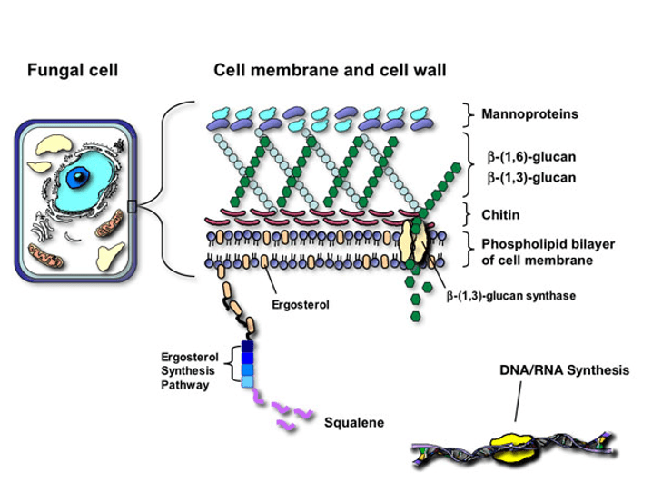
Intravenous amphotericin B is the current standard of care. Amphotericin B works by disrupting the selective permeability of plasma membranes. It works by binding to ergosterol (the sterol in fungi cell membranes). Though not a fungi, N. fowleri has ergosterol in its cell membrane. Hence, Amphotericin B, which is normally an antifungal medication, can also be used as an antiprotozoal medication for otherwise-untreatable diseases caused by certain protozoa.
In this case, Amphotericin B binds to ergosterol in the cell membrane of N. fowleri, giving rise to a pore which allows potassium ions and other small molecules to leak out from the cell, ultimately leading to cytolysis. Unfortunately, having great results in the lab doesn’t always necessarily translate to a similar level of success in actual patients.

Amphotericin B is a ‘last-resort’ drug as it has high toxicity, and its use in actual patients are not as successful in actual patients compared to what initial lab experiments predict.
So, just bear this all of the above in mind before you decide to venture out into warm ponds, freshwater rivers and lakes for a swim, especially during the hot summer months.
Well, I’m kidding. The odds are that you’re probably going to be fine. Nevertheless, take caution. Don’t dunk your head in the water at a natural hot spring for goodness’ sake.
There have only been a few hundred cases of Naegleria fowleri infection worldwide since the parasite was documented from the 1960s. Naegleria fowleri is actually quite common, but its infection is rare. However, if you are infected, then the prognosis isn’t particularly optimistic (>95% death).
I hope you have learnt more about this interesting parasite from reading this article!
Yours faithfully,
Nic Loh
23 July 2019
References & Further Reading
1. https://abcnews.go.com/Health/warm-weather-stirs-brain-eating-amoeba-warning/story?id=24552241
2. https://www.webmd.com/brain/brain-eating-amoeba#1
3. https://www.sciencedirect.com/topics/neuroscience/olfactory-nerve
4. https://www.sciencedirect.com/topics/medicine-and-dentistry/naegleria-fowleri
5. https://slideplayer.com/slide/7225568/
6. http://humanphysiology.academy/Neurosciences%202015/Chapter%201/P.1.1.2%20Compound%20Action%20Potential.html
7. https://images.app.goo.gl/dTh1i8e9Hau92jkY9
8. http://www.cdipd.org/index.php/naegleriasis-global-impact