
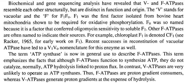
The world’s tiniest motor, ATP synthase, or F1FO-ATPase, stands at about 20 nm in height and 10 nm in diameter.
Look at the above artist’s impression of ATP synthase. What a beautiful image. This protein complex is something that many biology, biochemistry, health sciences students are required to learn about. Today, I wish to talk about ATP synthase as I find it quite fascinating.
To begin, let us resolve some issues regarding its nomenclature. ATP synthase belongs to a broader group known as ATPases. This might leave you wondering, as it did for me: “why does ATP synthase fall under this category? From its name alone, don’t ATPases couple the hydrolysis of ATP with endergonic reactions? In other words, don’t ATPases catalyse the dephosphorylation from ATP to ADP which would generate energy that would be harnessed by the ATPase to drive reactions which would otherwise not occur? ATP synthase isn’t hydrolysing ATP; it’s making ATP” It turns out that for most ATPases, ATP is hydrolysed; however, ATPases also include the inverse reaction to ATP hydrolysis, to wit, the phosphorylation of ADP with inorganic phosphate to give ATP.
It’s just like how the oxidation of succinyl-CoA to succinate is catalysed by succinyl-CoA synthetase. Even though succinyl-CoA isn’t the product in the forward reaction, the same enzyme is being used in both the forward and reverse reaction.
Under the umbrella term of ATPases, there are sub-categories: F-, V-, A-, P-, and E-ATPases. I will focus on F-ATPases (aka F1FO-ATPases), the most notable of which is ATP synthase. Unlike most of the other sub-categories of ATPases, F-ATPases catalyse the synthesis of ATP. A-ATPases work like F-ATPases too; it’s just that A-ATPases are found in Archaea. ATP synthase is sometimes known as Complex V.
“Nicholas, why is the F-ATPase called F1FO-ATPase? Shouldn’t it be named F1F0-ATPase? After all, isn’t the preceding number to 1 is obviously 0?” Let me explain. Oligomycin is an antibiotic that inhibits ATP synthase by blocking its proton channel through the Fo subunit. In my opinion, F0 has become commonly adopted by many authoritative textbooks and peer-reviewed articles for the following reasons:
1) The letter ‘o’ is visually similar to ‘0’. In classrooms or lecture theatres, where teachers and professors write about the Fo sub-unit by hand on the whiteboard, it might be impossible to determine if an ‘o’ or ‘0’ was written.
2) This is probably the biggest factor: ‘0’ precedes ‘1’. Meanwhile, ‘o’ is a letter while ‘1’ is a number.
3) There are many more symbols where ‘0’ is the subscript instead of ‘o’.
4) Many authoritative texts and teachers use F0 instead of Fo and when other people read or listen to them, they also do the same and adopt the convention. As such, the use of F0 is rapidly adopted.
5) There is little mention about this historical development in various resources.
This two-paragraph extract from a paper by McCarty (1992) elaborates on the situation.
Also of note is the fact that some older texts refer to ATP synthase as ATP synthetase.
According to the JCBN (Joint Commission on Biochemical Nomenclature)/NCIUBMB (Nomenclature Committee of the International Union of Biochemistry and Molecular Biology) newsletter in 1984, the word synthase can now be added to the name of any substance to signify an enzyme that catalyses a reaction producing that substance, i.e. its range is extended to include enzymes catalysing reactions that involve hydrolysis of a nucleoside triphosphate, which previously could not be called synthases. In addition, “synthetase” is now synonymous with “ligase”.
In view of this update, it is thus more precise to refer to Complex V as ATP synthase instead of ATP synthetase. I also want to talk about the theoretical or experimentally-determined values regarding how many ATPs are produced for each molecule of NADH or FADH2 that enters the mitochondrial electron transport chain. The oldest values are: each NADH and FADH2 molecule leads to the production of 3 ATP and 2 ATP respectively.
However, we also see values such as 2.5 ATP and 1.5 ATP for each molecule of NADH or FADH2 respectively. Some modern texts even state that each NADH leads to 2.7 ATP being produced and 1.6 ATP/FADH2. Which is it?
To understand how we got into this mess, we must understand the relevant historical development. In the 1960s, ATP was known to be the energy currency of life, but the mechanism by which ATP was generated in the mitochondrial matrix was assumed to be substrate-level phosphorylation. After all, substrate-level phosphorylation was seen in glycolysis, and the Krebs cycle where succinyl-CoA is oxidised to succinate, while dephosphorylating GTP to GDP, which in turn phosphorylates ADP to ATP.
A scientist named Peter D. Mitchell realised that the movement of ions across an electrochemical potential difference could provide the energy needed to produce ATP. His hypothesis was actually derived from information that was already well known in the 1960s. He knew that living cells had a membrane potential; interior negative to the environment. The movement of charged ions across a membrane is thus affected by the electrical forces (the attraction of positive to negative charges). Their movement is also affected by the chemical potential, where substances diffuse from regions of higher concentration. He went on to show that ATP synthesis was coupled to this electrochemical gradient.
But of course, as with many great scientists who came up with seemingly absurd hypotheses, I’m sure he was mocked and ridiculed for this. Here is a quote from a biography of Mitchell: “It is a striking fact that, at the time Mitchell proposed his theory, there was not a shred of experimental evidence in its favour.” When Mitchell died in 1992, the following obituaries were published:
From The Times, a British daily national newspaper, on 15 April 1992: “His hypothesis met, initially, with an almost derisory reception. Few of the workers in the field had the necessary background knowledge of electrochemistry to appreciate the proposals and they spent many years searching for a chemical intermediate, which Mitchell argued did not exist.”
From The Independent, established in 1986 as a politically independent national morning newspaper published in London, on 16 April 1992: ‘For 20 years Mitchell was ridiculed, and Jennifer Moyle was his only professional supporter’.
Mitchell’s contemporaries have been criticised for their hostility towards Mitchell’s chemiosmotic hypothesis. Such criticism may be misplaced, though. For example, the elegance of the hypothesis was immediately recognised by some. In a review published in 1962, Lehninger (yes, this is the same Lehninger as the one who authored the renowned biochemistry textbook) gave a sympathetic summary of the chemiosmotic hypothesis, and Mitchell was invited by his peers to present his hypothesis in seminars at various European universities.
Anyway, the chemiosmotic hypothesis eventually became the leading hypothesis. The brilliance of the chemiosmotic coupling was that it allows nonintegral stoichiometries of oxygen consumption and ATP synthesis. Let me explain.
Before the chemiosmotic model for oxidative phosphorylation was accepted, the assumption was that the overall reaction equation would take the following form because everyone thought that it would be substrate-level phosphorylation:
xADP + xPi + NADH + H+ + 0.5O2 —> H2O + NAD+ + xATP
where the value of ‘x’ would be an integer. When intact mitochondria are suspended in solution with a oxidisable substrate such as NADH or succinate, along with addition of oxygen, ATP synthesis is readily measurable, as is the decrease in oxygen. In theory, this should allow ‘x’ to be calculated. However, measurement of ‘x’ is complicated by the fact that 1) in intact mitochondria, any ATP that is produced is used by the mitochondria in other reactions in the matrix. Also, 2) oxygen is also used in reactions unrelated to oxidative phosphorylation. Hence the discrepancy due to these two factors need to be taken into account. Most experiments, after the necessary corrections, have determined x to be between 2 and 3 when 1 mole of NADH is used as the oxidisable substrate.
Given the assumption that x was to be an integer, most experimenters agreed that the x should be 3 and 2 for NADH and succinate respectively. Just like that, these values have been used for years in peer-reviewed articles and textbooks.
With the chemiosmotic model involving the coupling of ATP synthesis (involving phosphorylation) and electron transfer (involving oxidation), there was no longer a theoretical requirement for x to be an integer. The measurement of proton fluxes (how many protons are pumped through the various complexes) is quite complicated but basically, the experimental consensus is that for each pair of electrons, 10 protons and 6 protons are pumped into the intermembrane space for one molecule of NADH and succinate respectively. For flow back into the matrix from the IMS, the most widely accepted value is that 4 protons are required to drive the synthesis of 1 ATP.
Since 1 NADH leads to 10 protons being pumped from the matrix into the IMS, and 4 protons are required for synthesis of 1 ATP, it follows that x = 10/4 = 2.5 for NADH. Thus, 1 NADH leads to the production of 2.5 ATP. It also follows that since 1 succinate molecule leads to 6 protons being pumped from the matrix into the IMS, then x = 6/4 = 1.5 for succinate. Thus, 1 succinate leads to the production of 1.5 ATP.
Since 1 NADH leads to 10 protons being pumped from the matrix into the IMS, and 4 protons are required for synthesis of 1 ATP, it follows that x = 10/4 = 2.5 for NADH. Thus, 1 NADH leads to the production of 2.5 ATP. It also follows that since 1 succinate molecule leads to 6 protons being pumped from the matrix into the IMS, then x = 6/4 = 1.5 for succinate. Thus, 1 succinate leads to the production of 1.5 ATP.

You might have noticed that I talked so much about succinate. What happened to FADH2? Here’s the brilliance of the mitochondrion. The TCA Cycle takes place in the matrix, and recall that one of the steps of the TCA involves the oxidation of succinate to fumarate. Now, this oxidation actually occurs at Complex II of the mETC. The oxidation of succinate to fumarate simultaneously reduces FAD to FADH2. FAD is a prosthetic group, a covalently-attached cofactor that is found within Complex II. FADH2 is then oxidised back to FAD when it reduces Q to QH2. QH2 then shuttles to Complex III. This is how electrons are carried from the TCA, through II to III, etc! Amazing, isn’t it? Thus, it could be alternatively said that 1 FADH2 leads to 1.5 ATP.
We could use the same logic and apply it to NADH, but then NADH could come from so many places. Just imagine. 1 isocitrate produces 2.5 ATP; 1 α-ketoglutarate produces 2.5 ATP; 1 malate produces 2.5 ATP. This isn’t practical at all. From the TCA cycle alone, NADH is produced in 3 instances:
1) the oxidation of isocitrate to α-ketoglutarate simultaneously reduces NAD+ to NADH
2) the oxidation of α-ketoglutarate to succinyl-CoA simultaneously reduces NAD+ to NADH
3) the oxidation of malate to oxaloacetate simultaneously reduces NAD+ to NADH
NADH can also come from another reaction within the mitochondrial matrix which is not, strictly speaking, part of the TCA. Firstly, consider the earlier oxidation of α-ketoglutarate to succinyl-CoA, where this reaction is catalysed by α-ketoglutarate dehydrogenase complex. α-ketoglutarate (5 carbons) is oxidised to succinyl-CoA (4 carbons). What happened to that one carbon? Well, it escaped as CO2. Look at the product of the reaction too. A “CoA” is added. Also, an NADH is produced. The enzyme is a dehydrogenase complex.
Does this ring a bell?
If you thought of the oxidation of pyruvate to Acetyl-CoA, then CONGRATULATIONS, you got it!
The thing you must understand is this: α-ketoglutarate dehydrogenase complex is virtually identical to pyruvate dehydrogenase complex. Within the matrix, the oxidation of pyruvate (3 carbons) to Acetyl-CoA (2 carbons) produce CO2, brings about the addition of a “CoA”, and lastly, reduces NAD+ to NADH.
In addition, NADH can be shuttled from the cytosol into the matrix to be used as substrate at Complex I.
Let’s go back and talk about the proton flux where protons move from the IMS into the matrix. How does it work? ATP synthase consists of two parts: Fo and F1. Fo has a number of sub-units and the number of sub-units is equal to the number of protons that move through Fo. In animal mitochondria, n = 8 (it varies, depending on the organism; in E. coli, n = 10). This means that 8 protons move through Fo in one revolution of Fo. Also consider the F1 portion, where one revolution of F1 produces 3 ATP. Since the synthesis of each ATP molecule requires the transfer of 1 Pi into the matrix, at a cost of 1 proton for each Pi transfer, that means that 3 additional protons are required for the synthesis of 3 ATP. This means that 8 + 3 = 11 protons are needed for the synthesis of 3 ATP. Remember that this is animal mitochondria that we are talking about. 11/3 gives 3.7 protons/ATP for ATP synthesis in animals. When we use the values of 10 protons/NADH and 6 protons/succinate or FADH2, we get: 10/3.7 = 2.7 ATP/NADH and 6/3.7 = 1.6 ATP/succinate or FADH2 in animals.
Then you might be wondering: how does this relate to 2.5 ATP/NADH and 1.5 ATP/NADH respectively? Recall that the experimental consensus was 4 protons/ATP. Now, what organisms were used to obtain that value? That is the crux of the matter. In the previous paragraph, I mentioned that the number of Fo sub-units vary depending on the organism. This totally changes everything. For example, in the case of E. coli andyeast, where n = 10, the values you get will be 2.3 ATP/NADH and 1.4 ATP/succinate or FADH2, if you follow the same calculation process in the previous paragraph.
These above calculated values of x (aka P/O ratio) define a range that includes the experimental values of 2.5 ATP/NADH and 1.5 ATP/FADH2. Thus, 2.5 and 1.5 are ubiquitous in biochemistry textbooks. Yet, for simplicity, some still choose to use 3 ATP/NADH and 2 ATP/FADH2.
I hope this blog post has perhaps allow you to understand why I find ATP synthase so fascinating. It is a pity that a lot of the above background information which includes highly interesting historical development isn’t discussed enough in textbooks, classrooms, and lecture theatres because of a lack of time, resources, etc.
Yours faithfully,
Nic Loh
14 May 2019
References for your further reading pleasure
1. Lehninger. Principles of Biochemistry. 6e.
2. https://www.qmul.ac.uk/sbcs/iubmb/newsletter/misc/synthase.html
3. Kagawa Y & Racker E 1966, “Partial resolution of the enzymes catalyzing oxidative phosphorylation. 8. Properties of a factor conferring oligomycin sensitivity on mitochondrial adenosine triphosphatase”. The Journal of Biological Chemistry. 241 (10): 2461–6.
4. Mccarty RE 1992. “A plant biochemist’s view of H+-ATPases and ATP synthases”. The Journal of Experimental Biology. 172 (Pt 1): 431–441.
5. https://iubmb.org/biochemical-nomenclature/
6. https://en.wikipedia.org/wiki/Peter_D._Mitchell#Chemiosmotic_hypothesis
7. https://royalsocietypublishing.org/doi/pdf/10.1098/rsbm.1994.0040
8. https://en.wikipedia.org/wiki/ATP_synthase#F1_region
9. https://atlasofscience.org/multiple-tasks-for-the-c-ring-of-the-f1fo-atp-synthase/
10. https://www.sciencedirect.com/topics/biochemistry-genetics-and-molecular-biology/f1-atpase

Leave a comment